Dental Radiology
It is an extremely important element of the diagnostic process, as abnormalities visible on radiographs are often not detectable in the clinical examination of the patient.
Radiology Office with Panoramic Radiographs
This type of imaging is used by the dentist both during the first consultation to diagnose the patient's condition and develop an individual treatment plan, as well as during its implementation and after completion, during follow-up visits.
MALO CLINIC Warsaw has its own diagnostic equipment, including an orthopantomograph (commonly referred to as panoramic X-ray) and a own computed tomography scanner.
In dentistry, we perform several types of X-rays:
-
Dental X-rays (point radiographs)
These are most commonly performed in dentistry. They are used for diagnostics before tooth extraction, before/after/during root canal treatment, to detect dental caries – especially on interproximal surfaces of teeth and inflammatory conditions of the periapical tissues. They are also used to assess the integrity of fillings, the condition of the marginal periodontium, detect tooth trauma, and abnormalities in tooth development or structure.
-
Bite-wing X-rays
These reveal the crowns of the upper and lower teeth along with the marginal periodontium, but the periapical areas are not visible.
-
Occlusal X-rays
These can be used to locate impacted or supernumerary teeth; in cases of trauma, to assess fractures of teeth and alveolar bones in the anterior part of the dental arch; for diagnostics of the floor of the mouth to detect salivary stones; to visualize cleft palate fissures. They are often performed in children to assess the area around the apices of the teeth.
-
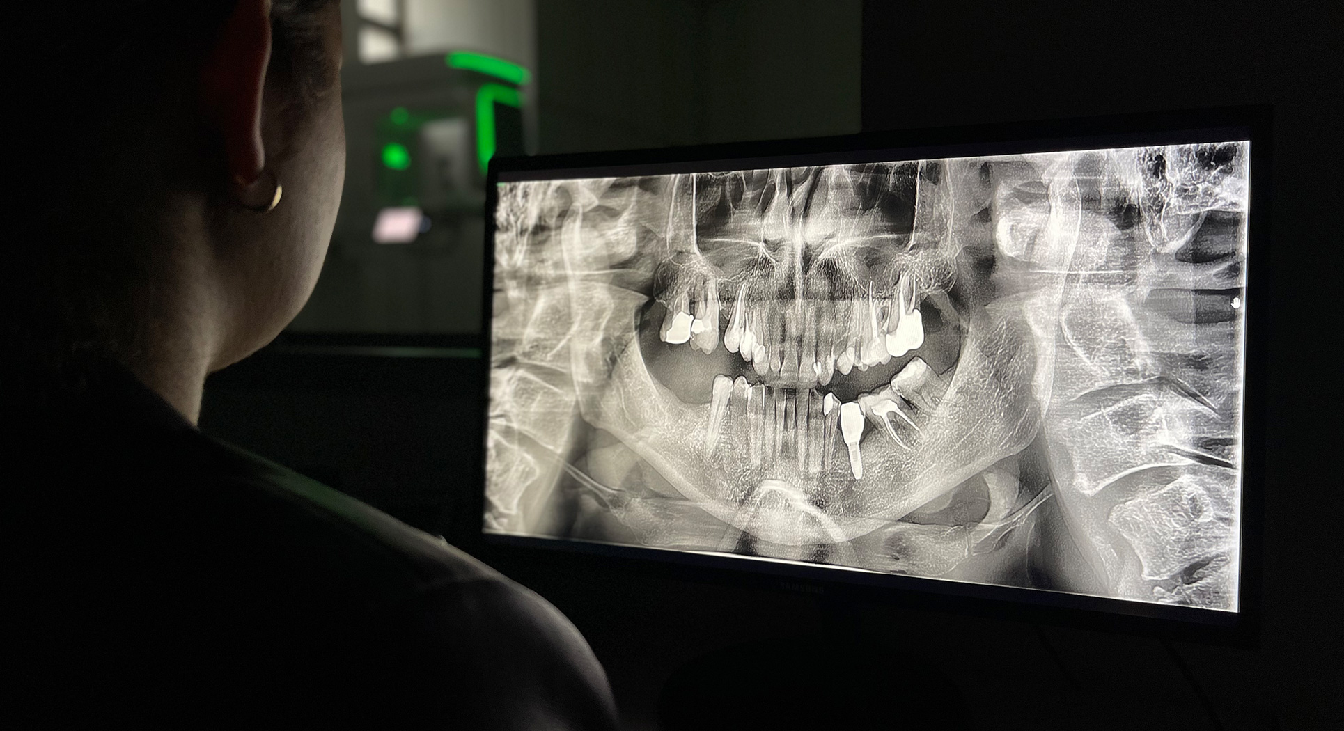
Panoramic X-rays
These allow visualization of both upper and lower teeth, surrounding tissues, as well as temporomandibular joints and sinuses. They help assess the presence of caries, roots left after extractions, the quality of fillings and root canal treatments, detect impacted teeth, estimate the stage of periodontal disease, and can detect asymptomatic cysts or even tumors. -
Cefalometric X-rays
These enable the diagnosis of malocclusions and planning of orthodontic treatment by performing angular and linear cranio-metric (inherited facial skeletal structure) and gnathometric (disorders of the masticatory apparatus) measurements. The visible outline of the soft tissues is used for facial profile analysis. -
Computed Tomography
Radiographs show 3D structures only in two dimensions, so to properly assess the width and height of the alveolar bone, bone quality, and the position of anatomical structures such as the maxillary sinus, nasal cavity, or nerve canals, a computed tomography scan is necessary. A 3D image is typically performed before starting implant treatment. This scan aids in selecting the most suitable implant dimensions, ensuring a safe and minimally invasive procedure. Tomography is also used in endodontics to locate additional root canals and to diagnose teeth with periapical changes and their extent.
Main tasks of dental X-rays:
- Diagnosing pathological changes in the mucosa, bone, and teeth
- Diagnosing temporomandibular joint pathology
- Assessing bone quality and quantity when planning implant surgery
- Assessing the location of nerve canals in complex extractions (e.g., impacted teeth)
- Planning orthodontic, prosthetic, and endodontic treatment
The use of digital technology enables the real-time transmission of X-ray images between the radiology office and the dental clinic, allowing the doctor to immediately review the images.